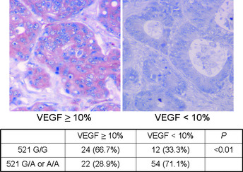
Figure 3.

Representative immunohistochemical staining patterns of vascular endothelial growth factor (VEGF). The staining results for VEGF were divided into two groups, <10% and more than 10% positive cells, according to the percentage of carcinoma cells showing specific immune‐reactivity.
