
Figure 1.
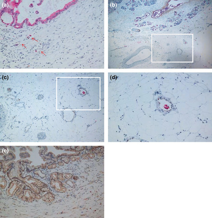
Cytokeratin 19 (CK19) and D2‐40 dual immunohistochemistry revealing lymphatic vessel (LV) invasion, peripancreatic plexus invasion, and lymph node metastasis. (A) High‐density area of LV in peripancreatic tissue. Most lymphatic vessels exhibited lumen‐like structures with tumor cells inside. Red arrows indicate lymphatic endothelial cells stained by D2‐40 and pancreatic cancer cells stained by CK19 (original magnification ×200). (B) Lymphatic vessels, blood vessels and nerve bundles showing CK19‐stained tumor cells inside the LV (original magnification ×40). (C) Staining of pancreatic tumor cells for CK19 (red) and peripancreatic LV for D2‐40 (brown‐yellow; original magnification ×100). Panel C shows the boxed area in (b) and panel D shows the boxed area in (c). (D) Tumor cells staining positive for CK19 (red) were found inside the peripancreatic LV (brown‐yellow; original magnification ×200). (E) Vascular endothelial growth factor‐C‐stained regions found in the plasma of tumor cells (brown‐yellow yellow; original magnification ×200).