
Figure 2.
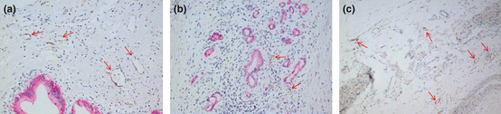
Cytokeratin 19 (CK19) and D2‐40 dual immunohistochemical analysis of lymphatic vessel density (LVD). (a) Peripancreatic tissue showing areas of high LVD. Most lymphatic vessels maintained lumen‐like structures (original magnification ×200). (b) Within pancreatic cancer tissues, low LVD was observed. The lymphatic vessels lost their structure due to the pressure created by the tumor (CK19 [red]‐stained pancreatic cancer cells in D2‐40 [brown‐yellow]‐stained lymphatic endothelial cells; original magnification ×200). (c) The peripancreatic nerve plexus region showing areas of high LVD. Most lymphatic vessels presented with enlarged lumen‐like structures. Arrows indicate lymphatic endothelial cells stained with D2‐40. (Original magnification ×100.)