
Figure 3.
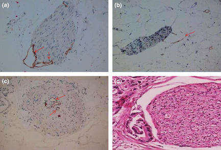
(a–c) Cytokeratin 19 (CK19) and D2‐40 dual immunohistochemical analysis of the distribution of lymphatic vessels (LV) in the peripancreatic plexus. (a) Peripancreatic LV accompanied by the peripancreatic plexus, showing close contact or invasion into the epineurium (original magnification ×200). (b) Peripancreatic LV in close contact or invading the epineurium. The LV are seen as semi‐open structures (original magnification ×200). (c) Peripancreatic LV directly entering the peripancreatic nerve bundle (original magnification ×200). (d) Invasion of the peripancreatic plexus showing infiltration of the epineurium and partial perineurium (H&E staining; original magnification ×100). Arrows indicate lymphatic endothelial cells stained by D2‐40.