Figure 1.

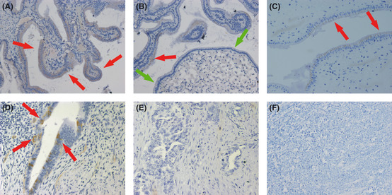
Expression of tissue factor pathway inhibitor‐2 (TFPI‐2) in gallbladder carcinoma (GBC) tissues, lymph metastasis, normal tissues, and gallbladder polyp (GBP) tissues by immunohistochemical staining (×200). The percentage of positive‐stained epithelial cells or cancer cells with >10% were considered as positive. Red arrow represents positive expression, and green arrow represents negative expression. (A) TFPI‐2 expression in normal tissues. TFPI‐2 staining was located in the cytoplasm of epithelial cells. (B) TFPI‐2 expression was detected in epithelial cells of normal tissue but not in those of GBP tissues. (C) TFPI‐2 expression was also detected in epithelial cells of GBP tissues. (D) TFPI‐2 expression was detected in cancer cells of GBC tissues. (E) TFPI‐2 expression was not detected in cancer cells of GBC tissues. (F) TFPI‐2 expression was not detected in cancer cells of lymph metastasis.
