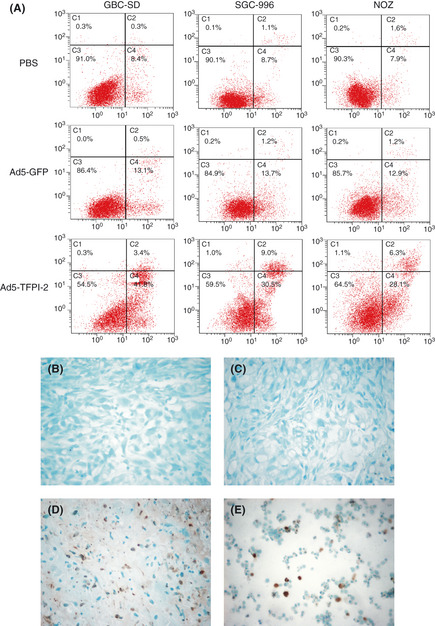
Figure 4.

Apoptosis in gallbladder carcinoma (GBC) cell lines and tissues was evaluated by Annexin V‐APC/PI assay (A) and terminal deoxynucleotidyl transferase‐mediated dUTP nick end labeling (TUNEL) assay (B,C,D), respectively. (A) Adenovirus‐mediated gene transfer of tissue factor pathway inhibitor‐2 (Ad5‐TFPI‐2) induced apoptosis in all three GBC cell lines (P = 0.000). Cell apoptosis was examined by using Annexin V and propidium iodide (PI) staining. X‐axis represents Annexin V fluorescence intensity and Y‐axis represents PI fluorescence intensity. Cells that stain positive for Annexin V and negative for PI are undergoing apoptosis (lower right). Cells that stain positive for both Annexin V and PI are either in the end stage of apoptosis, undergoing necrosis or already dead (upper right). (B) Phosphate‐buffered saline (PBS) group (×400). (C) Ad5‐GFP group. (D) Ad5‐TFPI‐2 group. (E) Positive control (a mixture of HL60 cells incubated with 0.5 μg/mL actinomycin D for 19 h to induce apoptosis, provided by Merck). A dark brown 3,3′‐diaminobenzidine tetrachloride (DAB) signal indicates positive staining while shades of blue to green signifies a non‐reactive cell. Counterstaining with methyl green aids in the morphological evaluation and characterization of normal and apoptotic cells.
