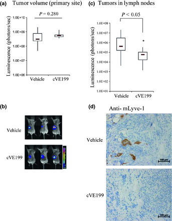
Figure 5.

In vivo effect of cVE199 against vascular endothelial growth factor (VEGF)‐D expressing neuroblastoma model. (a and b) Antitumor effect of cVE199 against SK‐N‐DZ primary lesion was using a bioimaging technique. Mice bearing SK‐N‐DZ were give 10 mg/kg of cVE199 twice a week for 2 weeks. Tumor size was measured by administration of luciferase substrate and detection of the photons. Data are shown by a box‐and‐whisker plot. Statistical tests were performed using the Wilcoxon test. (c) Anti‐metastasis activity of cVE199 against SK‐N‐DZ was evaluated by detection of photons from lysed lymph nodes. Data are shown by a box‐and‐whisker plot. Statistical tests were performed using the Wilcoxon test. (d) The presence of lymph vessels in the primary tumors of SK‐N‐DZ was analyzed by mLyve‐1 immunostaining. Formalin‐fixed paraffin‐embedded slides were stained with an anti‐mouse Lyve‐1 antibody using a Ventana automated immunostainer.
