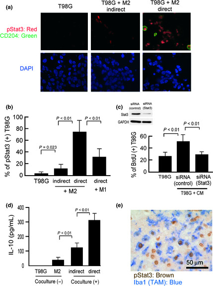
Figure 2.

Signal transducer and activator of transcription‐3 (Stat3) activation in cocultured cells. (a) T98G cells were cocultured with tumor‐cell supernatant‐stimulated M2 macrophages, and Stat3 activation was analyzed by double immunostaining of pStat3 (red) and CD204 (green). Blue indicates nuclear staining. Scale bar = 50 μm. (b) Following double immunostaining, 100 CD204− T98G cells were counted, and the percentage of pStat3+ cells was calculated (n = 3 or 4 for each group). (c) BrdU incorporation in conditional medium‐stimulated T98G cells was evaluated with or without Stat3 siRNA. (n = 3 for each group). Downregulation of Stat3 protein in T98G cells was also confirmed by Western blot analysis. (d) Interleukin (IL)‐10 production was evaluated as a marker of macrophage activation (n = 4 for each group). (e) Double immunostaining of activated Stat3 (brown) and Iba‐1 (marker of macrophages/microglia; blue) was carried out.
