
Figure 6.
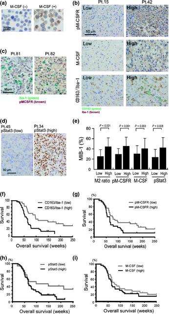
Immunohistochemical determination of phosphorylated macrophage‐colony stimulating factor receptor (pM‐CSFR), macrophage‐colony stimulating factor (M‐CSF), phosphorylated signal transducer and activator of transcription‐3 (pStat3), anti‐Ki‐67 (clone MIB‐1), and M2 ratios in human high grade gliomas. (a) TF‐1 culture cells were stimulated by M‐CSF for 5 min and activation of M‐CSFR was evaluated by immunostaining with anti‐pM‐CSFR. Strong activation of pM‐CSFR was detected on cell surfaces of M‐CSF‐stimulated TF‐1 cells. (b) Immunostaining of pM‐CSFR, M‐CSF, and double immunostaining of CD163 (green) and Iba‐1 (brown) were carried out. Results for patient (Pt.) no. 15 and no. 42 are shown. (c) By double immunostaining ofIba‐1 (green) and pM‐CSFR (brown), pM‐CSFR was detected in both tumor cells and macrophages. (d) Immunostaining of pStat3 was also carried out to evaluate the Stat3 activation in tumor cells. (e) M2 ratio, M‐CSFR activation, Stat3 activation, and M‐CSF expression were correlated with the MIB‐1 index. The Kaplan–Meier method was used to determine (f) M2 ratio, (g) M‐CSFR activation, (h) Stat3 activation and (i) M‐CSF expression.