
Figure 2.
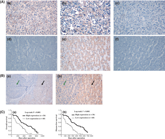
Immunohistochemistry of vacuole membrane protein 1 (Vmp1) expression in hepatocellular carcinoma (HCC) tissues and its prognostic implication. (A) Low expression of Vmp1 in HCC tissues (a–d) compared with high expression of Vmp1 in paracarcinomatous liver tissue (PCLT) (e) is shown; the negative control (f) is also included to show the specificity of the antibody. In these representative images, Vmp1 expression in the cytoplasm and membrane is scored as 3 + (a), 2 + (b), 1 + (c) and 0 (d) according to Shimizu criteria. Original magnification, ×400 (a–f). (B) Comparison of Vmp1 protein expression between HCC tissues (a) and PCLT (b). Original magnification, ×100 (a) and × 400 (b). Green arrows represent HCC and black arrows represent PCLT. (C) Kaplan–Meier survival curves of overall survival and disease‐free survival for the low Vmp1 expression group (scored as 0 and 1 +, n = 68) and the high Vmp1 expression group (scored as 2 + and 3 +, n = 56) based on the results of immunohistochemistry. The log‐rank test shows that HCC patients with low Vmp1 expression have lower overall survival (left) and disease‐free survival (right) than those with high expression of Vmp1.