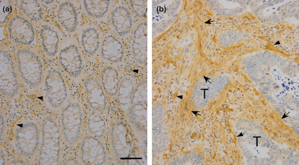
Figure 1.

Immunohistochemical analysis of angiomodulin (AGM) expressed in normal epithelial tissues and cancer tissues of colon. A single frozen section of a colon carcinoma (approximately 3 × 3 mm square size) was immunostained for AGM as described in Materials and Methods. (a) Normal glandular tissue, (b) stroma adjacent to tumor cells. Arrowheads, AGM‐positive micrrovessels; arrows, AGM‐positive stroma. Scale bars, 50 μm. When non‐immune mouse IgG was used as a negative control instead of the first antibody, no staining was found in the same conditions.
