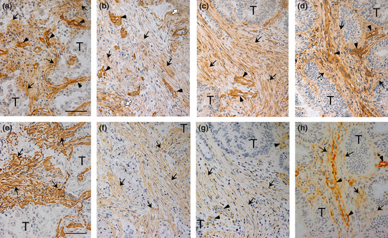
Figure 3.

Two close sections containing tumor cells (T) of lung adenocarcinoma (a,e), lung squamous cell carcinoma (b,f), colon adenocarcinoma (c,g) and uterus adenocarcinoma (d,h) were immunostained for angiomodulin (AGM) (a–d) and α‐smooth muscle actin (α‐SMA) (e–h). The colon cancer sections were derived from a different cancer sample from Figure 1. Arrows, positive signals for AGM or α‐SMA in stroma; arrowheads, AGM‐ or α‐SMA‐positive blood vessels; open arrowheads, AGM‐positive invading tumor cells in (b). Scale bars, 50 μm.
