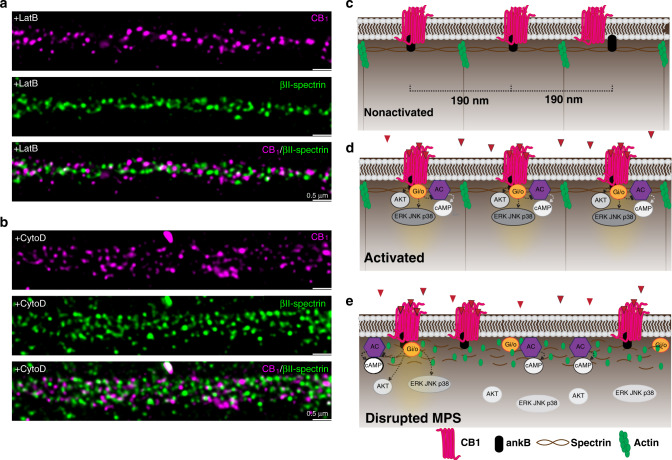
Fig. 5. Schematic illustration showing CB1 forming dynamic peri-periodic hotspots to increase signaling efficiency.
a, b Two-color STED images of CB1 (magenta) and βII-spectrin (green) in neurons treated with LatB (a) and CytoD (b). N = 3 biological replicates. c Without ligand binding, MPS sets the range for CB1 distribution. d Upon ligand binding, active CB1 are recruited to the MPS and become more periodic, making downstream AKT and ERK signaling more effective. e MPS degradation leads to less strong periodic clusters of CB1 and thus less downstream signaling.

