
Figure 2.
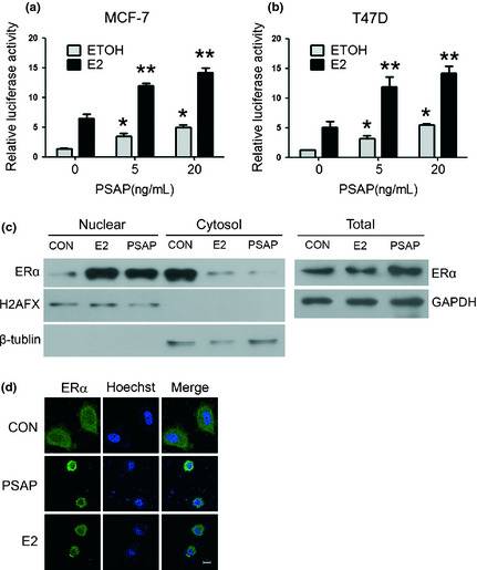
Prosaposin (PSAP) increased the transcriptional activity of estrogen receptor alpha (ERα). (a,b) MCF‐7 and T47D cells were transfected with pRL and 3×ERE‐LUC plasmids. After 24 h, cells were incubated with indicated concentrations of PSAP protein and ethyl alcohol (ETOH) or 100 nM E2, and harvested after another 24 h. Luciferase activity was measured and normalized to Renilla luciferase activity. (*P < 0.05 compared with ETOH control; **P < 0.05 compared with E2 control) (c) MCF‐7 cells were treated in the presence or absence of PSAP or E2 in serum‐ and PR‐free Dulbecco's modified eagle medium (DMEM) for 3 h. Nuclear and cytoplasmic extracts were prepared using a nuclear extraction kit and whole cell lysates were prepared from parallel tissue culture plates. Protein sample was subjected to sodium dodecyl sulfate‐polyacrylamide gel electrophoresis (SDS‐PAGE). Immunoblotting was performed using the anti‐ERα antibody. Glyceraldehyde 3‐phosphate dehydrogenase (GAPDH), β‐tublin and H2AFX were used as loading controls of whole cell lysates, cytoplasmic and nuclear extracts respectively. (d) Immunofluorescence assay was performed in MCF‐7 cells. The subcellular localization of ERα (green) and nuclear (blue) are shown. Scale bar, 10 μm.