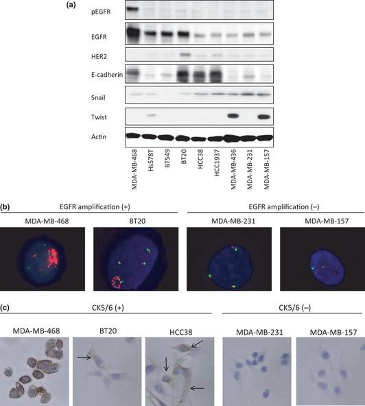
Figure 3.

Determination of breast cancer subtypes using basal markers and stem cell‐like characteristics. (a) Protein expressions of p‐EGFR, EGFR, HER2, E‐cadherin, Snail, Twist, and β‐actin in nine triple‐negative breast cancer (TNBC) cell lines. Ten micrograms of protein were prepared from the indicated cell lines at 60–70% confluence. The cell lines are arranged in order of decreasing sensitivity to everolimus, from left to right. β‐Actin was used as a loading control. (b) Epidermal growth factor receptor (EGFR) gene fluorescence in situ hybridization (FISH) analysis. MDA‐MB‐468 and BT20 showed the gene amplification of EGFR. The other seven TNBC cell lines did not exhibit the gene amplification of EGFR. Two positive cell lines and two negative cell lines are shown. (c) Immunohistochemical analysis of CK5/6. MDA‐MB‐468, BT20, and HCC38 were positive for CK5/6, and the other six cell lines were negative. Three positive cell lines and two negative cell lines are shown. Cell membranes were stained by the CK 5/6 antibody in all MDA‐MB‐468 cells and in some BT20 and HCC38 cells (indicated by arrows).
