Figure 1.

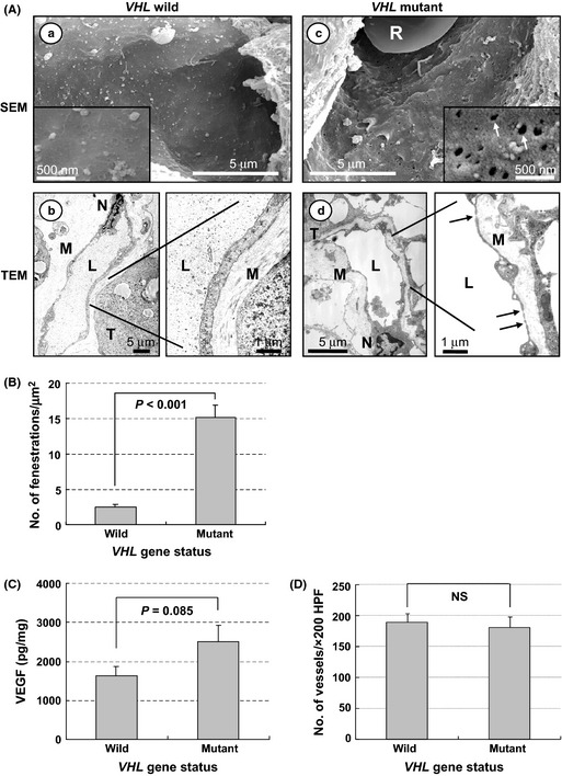
Phenotypes of tumor capillaries and VHL status in sporadic clear cell renal cell carcinomas (CC‐RCCs). (A) Electron micrographs of tumor capillaries in VHL wild‐type CC‐RCC (a,b) and VHL mutated CC‐RCC (c,d). Representative data are shown. Capillaries from tumors with wild‐type VHL show thick endothelium with few endothelial fenestrations (EFS); in contrast, those with mutant VHL show attenuated endothelium with abundant EFs (arrows). Inset shows a high magnification view (×50 000). L, capillary lumen; M, interstitial matrix; N, endothelial cell nucleus; R, red blood cell; SEM, scanning electron microscopy; T, tumor cell; TEM, transmission electron microscopy. (B) Quantitation of EFs in tumor capillaries with or without VHL mutation. Numbers of EFs were calculated per square micrometer. Columns, mean; bars, SE. (C) Amount of vascular endothelial growth factor (VEGF) in tumors with or without VHL mutation. Whole protein was extracted from tumors and analyzed by ELISA. Data are presented as pg VEGF protein/1 mg total protein. Columns, mean; bars, SE. (D) Microvessel density in tumors with or without VHL mutation. Columns, mean; bars, SE. HPF, high power field; NS, not significant.
