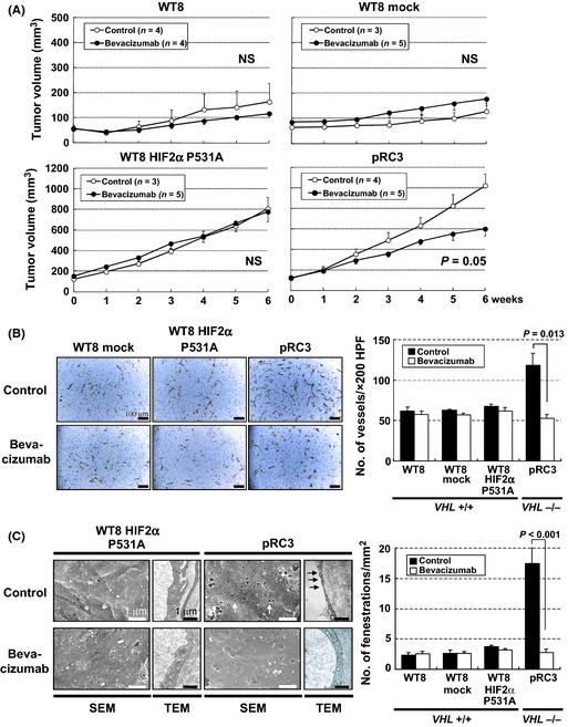
Figure 4.

Effects of bevacizumab treatment in renal cell carcinoma xenografts from 786‐O subclones with different statuses of VHL and hypoxia‐inducible factor (HIF) activity. (A) Antitumor effects of bevacizumab in renal cell carcinoma tumors in nude mice. Each time point represents the mean ± SE of fold of tumor volume in each group. The difference in tumor size between the treatment mice and controls was statistically significant in pVHL‐defective pRC3 xenografts (P = 0.05, using two‐way repeated anova). (B) Effects of bevacizumab treatment on microvessel density. Left, CD31 staining of xenograft tumors treated with bevacizumab (bottom) or vehicle (top). Scale bars = 100 μm (×200). Right, significant reduction of microvessel density was observed only in pRC3 xenografts. Columns, mean; bars, SE. (C) Effects of bevacizumab treatment on endothelial fenestrations. Left, Scanning electron micrographs (SEM) and transmission electron micrographs (TEM) of tumor capillaries with bevacizumab treatment (bottom) or vehicle (top). Scale bars = 1 μm (SEM, ×15 000; TEM, ×20 000). Right, reduction in the number of endothelial fenestrations in capillaries was detected only in pRC3 xenograft treated with bevacizumab. Columns, mean; bars, SE.
