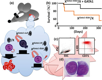
Figure 2.

Multi‐step leukemogenesis caused by the dysregulation of Gata1 gene expression. (a) There are two types of erythroid progenitor cells in females heterozygous for the Gata1 gene mutation due to the random inactivation of the X chromosome. Erythroid progenitor cells with the activated wild‐type allele undergo terminal maturation, whereas those with the inactivated wild‐type allele fail to differentiate and continue to proliferate. Immature cells with the activated Gata1‐null allele die by apoptosis due to the absence of GATA1. In contrast, a low level of GATA1 expression elongates the lifespan of the progenitor cells expressing either the activated Gata1‐IEdel or Gata1.05 allele. Consequently, those cells may acquire additional genetic event(s). (b) Survival curves of Gata1‐IEdel heterozygous females with (black line; n = 35) or without (red line; n = 10) the G1HRD‐GATA1 transgene. (c) A typical flow cytometry dot plot showing leukemic cell population positive for c‐Kit and CD71 (right panel). Left panel depicts a graph for side‐scatter (SSC) versus forward‐scatter (FSC). Leukemic cells in peripheral blood are gated in a red circle. (d) Leukemic cells in bone marrow of Gata1‐IEdel heterozygous females are morphologically similar to proerythroblasts.
