
Figure 1.
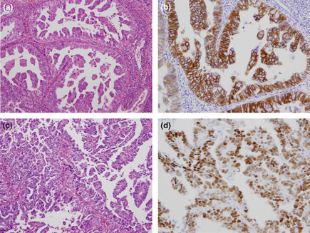
Histopathological presentation of uterine papillary serous carcinoma. (a,c) Hematoxylin and eosin staining, original magnification ×200. (b) Immunohistochemical staining showing overexpression of human epidermal growth factor receptor type 2 (HER2; score 3+). Original magnification ×200. (d) Immunohistochemical staining showing expression of the estrogen receptor (Allred score 5 + 3 = 8). Original magnification ×200.