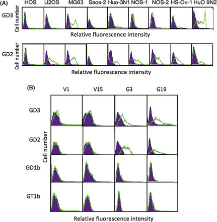
Figure 1.

Expression of GD3 and GD2 gangliosides on osteosarcoma cell lines and generation of GD3/GD2‐expressing lines from the human osteosarcoma HOS cell line. The expression of gangliosides was analyzed by flow cytometry. (A) Results of GD3 and GD2 expression. (B) Flow cytometry of GD3/GD2 expression on two controls (V1, V15) and two GD3 synthase–cDNA transfectant cells (G3, G19) was carried out with mAb R24, mAb 220‐51, and FITC‐labeled secondary antibody. Expression of GD1b and GT1b, located downstream of GD3 and GD2, was also analyzed by mAb 370 and mAb 549, respectively.
