Figure 1.

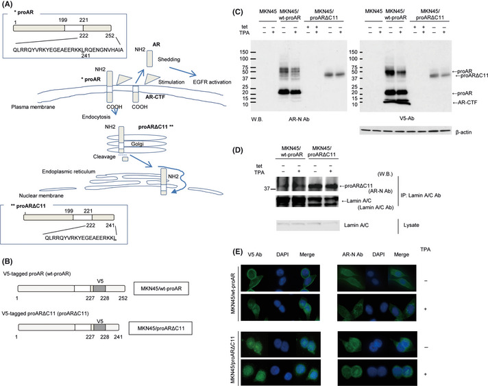
(A) Amphiregulin (AR) is expressed on the plasma membrane as proAR, consisting of a predicted 252 amino acids. ProAR primarily localizes at the plasma membrane. Stimulation induces partial proAR‐ectodomain shedding, resulting in production of soluble AR and the carboxyl‐terminal fragment of proAR (AR‐CTF). In addition, stimulation causes endocytosis of the residual proAR. During translocation to the nucleus, proAR is truncated to the 11 amino acids at the C‐terminus (proARΔC11), and proARΔC11 is targeted to the endoplasmic reticulum (ER). ER‐localized proARΔC11 subsequently diffuses or is actively transported to the nucleus. (B) Schematic presentation of V5‐tag inserted proAR and its deletion mutants. V5‐tagged proAR protein was termed wt‐proAR, and V5‐tagged proARΔC11 protein was proARΔC11. MKN45 lines transfected with the tet‐off system for consistent and conditional induction of wt‐proAR and proARΔC11 were named “MKN45/wt‐proAR” and “MKN45/proARΔC11,” respectively. (C) Western blot analysis of proAR and AR‐CTF expression in MKN45, MKN45/wt‐proAR and MKN45/proARΔC11. Each lane contained 100 μg of protein. The concentration of 12‐0‐tertadecanoylphorbor‐13‐acetate (TPA) was 200 nmol/L to induce ectodomain shedding. We used anti‐V5 antibody (V5 Ab) to recognize the cytoplasmic region of wt‐proAR and proARΔC11, and anti‐AR antibody (AR‐N Ab) to recognize the proAR ectodomain. Loading control comprised β‐actin. (D) Immunoprecipitation and western blot analysis of MKN45/wt‐proAR and MKN45/proARΔC11. The concentration of TPA was 200 nmol/L. Cell lysates were immunoprecipitated with anti‐lamin A/C antibody (Lamin A/C Ab), and precipitated proteins were analyzed by western blotting using AR‐N Ab and Lamin A/C Ab. The cell lysates were analyzed using Lamin A/C Ab. (E) Intracellular localization of proAR after TPA‐inducible processing in MKN45/wt‐proAR and MKN45/proARΔC11 under immunofluorescence microscopy. The concentration of TPA was 200 nmol/L. Nucleus stained blue with DAPI, AR stained green with AR‐N Ab and AR‐CTF stained green with V5 Ab.
