Figure 3.

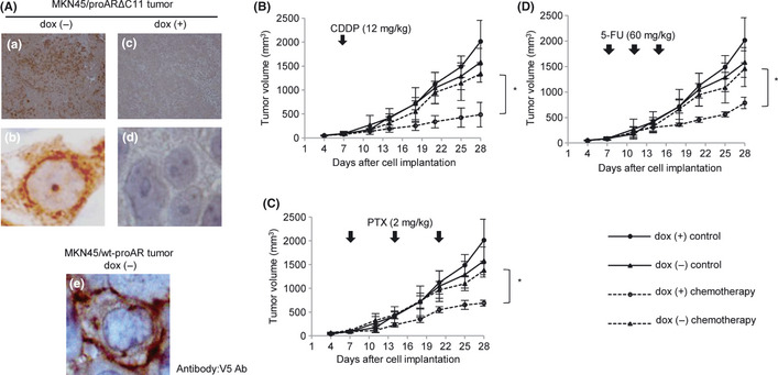
(A) Expression of proARΔC11 and wt‐proAR in xenograft tumor. We used V5 Ab to recognize transfected proARΔC11 and wt‐proAR. Immunohistochemical staining of proARΔC11 xenograft tumor without doxycycline (dox [−]) (a, ×100; b, ×1000). Immunohistochemical staining of proARΔC11 xenograft tumor with doxycycline (dox [+]) (c, ×100; d, ×1000). Immunohistochemical staining of wt‐proAR xenograft tumor dox (−) with large magnification (e, ×1000). (B–D) Subcutaneous MKN45/proARΔC11 tumor growth curves for nude mice after treatment with any anti‐cancer drugs (B, Cisplatin [CDDP]; C, paclitaxel [PTX]; D, 5‐fluorouracil [5‐FU]). ●, no chemotherapy dox (+); ▲, no chemotherapy dox (−); ○, each anti‐cancer drug dox (+); △, each anti‐cancer drug dox (−). Data are shown as means of four independent experiments. Bars, SD. (B) Intraperitoneal administration of 12 mg/kg of CDDP once on day 7. (C) Intraperitoneal administration of 2 mg/kg of PTX once weekly for 3 weeks. (D) Intraperitoneal administration of 60 mg/kg of 5‐FU once on days 7, 11 and 15. *, P < 0.05: treatment by each anti‐cancer drug (CDDP, PTX, 5‐FU) in dox (+) vs treatment by each anti‐cancer drug in dox (−).
