Figure 4.

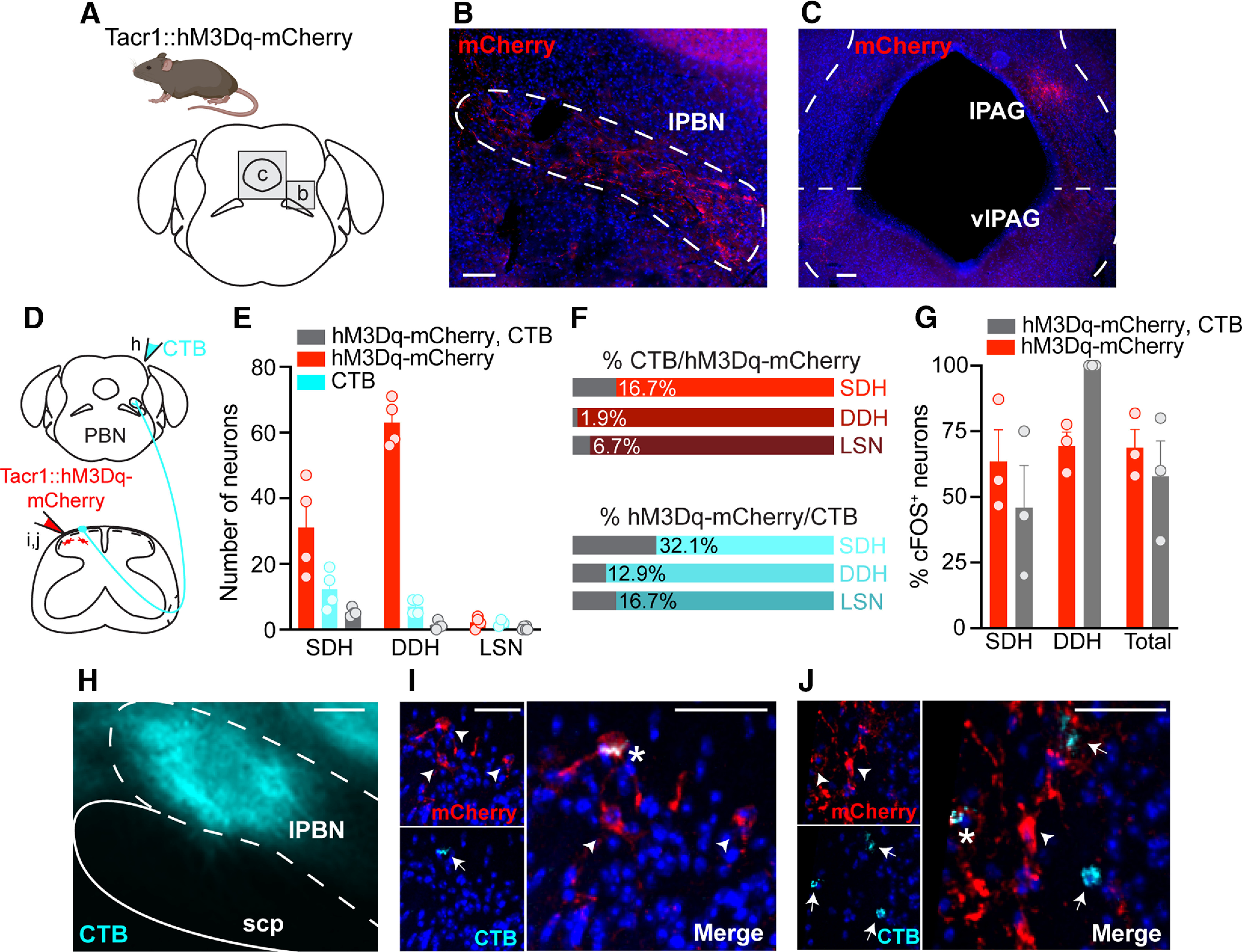
Tacr1CreER spinal neurons are predominately interneurons. A, The brains of Tacr1::hM3Dq-mCherry neurons were collected and sectioned coronally to evaluate whether Tacr1CreER captured spinal projection neurons. Representative immunostained coronal brain sections showing hM3Dq-mCherry processes (red) within the (B) lPBN, as well as the (C) lPAG and vlPAG contralateral to intraspinal viral injection. Scale bars: 50 μm. D, Experimental strategy to determine the relative proportion of Tacr1::hM3Dq-mCherry spinal projection neurons versus interneurons. The retrograde tracer CTB was injected into the contralateral lPBN of Tacr1::hM3Dq-mcherry mice to distinguish spinoparabrachial neurons (CTB and virally-mediated hM3Dq-mCherry expression) from interneurons (virally-mediated hM3Dq-mCherry expression only). E, Quantification of the total number of neurons labeled by hM3Dq-mCherry, CTB, or both within the SDH, DDH, and LSN of Tacr1::hM3Dq-mCherry mice, revealing that the majority of Tacr1CreER spinal neurons are local interneurons (n = 4 mice, 5 sections/mouse). F, Percentage of (top) total hM3Dq-mCherry neurons that were dual-labeled with CTB and (bottom) total CTB neurons that were dual-labeled with hM3Dq-mCherry within the SDH, DDH, and LSN, based off of number of neurons presented in E. G, Quantification of the percentage of cFOS+ hM3Dq-mCherry and hM3Dq-mCherry, CTB neurons across the dorsal horn shows equal activation following CNO administration (Student's t test, p = 0.513, t = 7.18, df = 4, n = 3 mice). Data are the same as those reported in Figure 3K–L but were reanalyzed to evaluate cFOS immunoreactivity in interneurons versus spinoparabrachial neurons of Tacr1::hM3Dq-mCherry mice. cFOS expression was detected within a single LSN in one mouse (Fig. 3L), and thus LSN neurons are not included here. H, Representative image of a targeted CTB injection (cyan) into the lPBN. Scale bar: 50 μm. I, J, Representative IHC images of lumbar spinal cord sections demonstrating the small extent of colocalization between hM3Dq-mcherry (red) and CTB (cyan). Scale bars, 10 μm. Arrowhead or arrow, labeled neuron; Asterisk, dual-labeled neuron. lPBN, lateral parabrachial nucleus; scp, superior cerebellar peduncle; lPAG, lateral PAG; vlPAG, ventrolateral PAG. SDH, superficial dorsal horn; DDH, deeper dorsal horn; LSN, lateral spinal nucleus. E, G, Data are shown as mean ± SEM, with open circles representing individual mice.
