Figure 5.

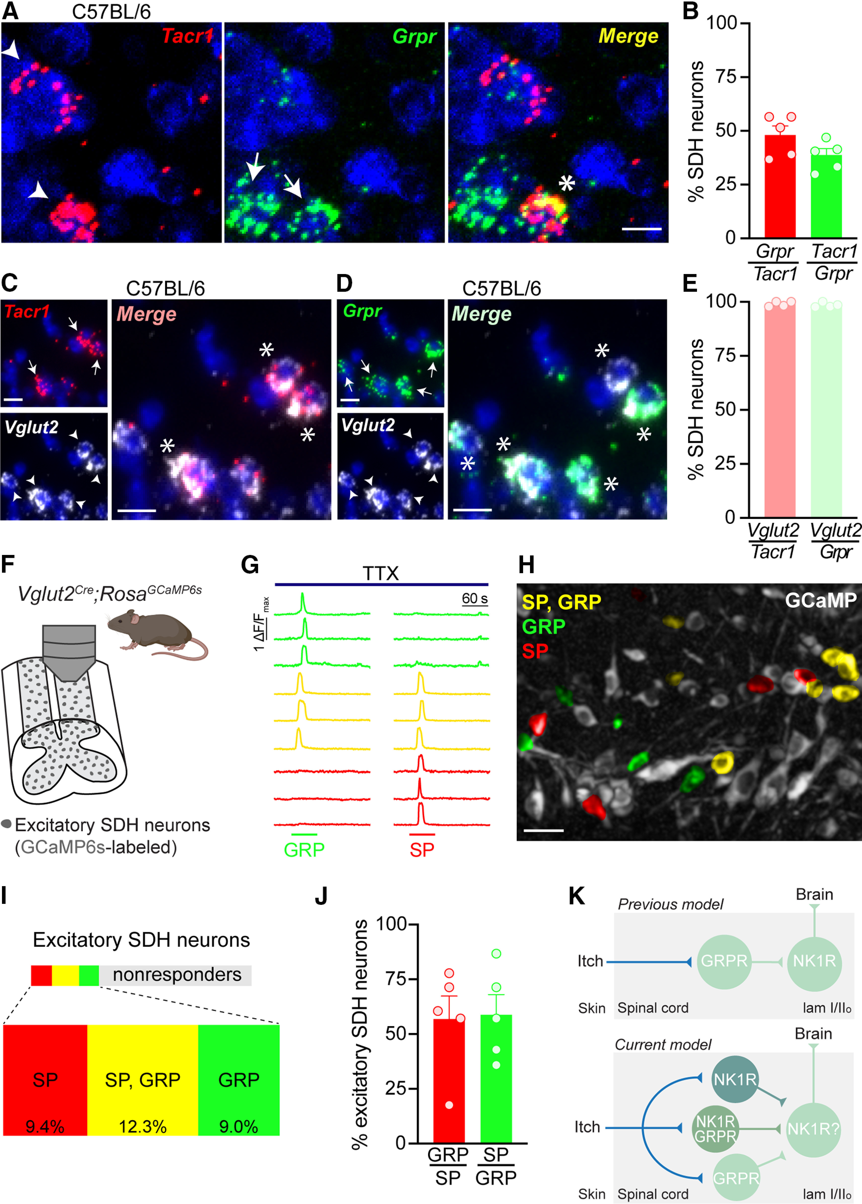
NK1R neurons in the SDH are a subset of GRPR interneurons. A, Representative images of dual FISH for Tacr1 (red) and Grpr (green) performed on lumbar spinal cord sections from C57BL/6 mice. Scale bar: 10 μm. B, Percentage of SDH neurons that coexpress Tacr1 and Grpr in C57BL/6 mice (n = 5 mice). Representative images of dual FISH evaluating the expression of the excitatory neuronal marker Vglut2 (white) in (C) Tacr1-expressing and (D) Grpr-expressing SDH neurons in C57BL/6 mice. Scale bars: 10 μm. E, Percentage of Tacr1 and Grpr SDH neurons that coexpress Vglut2 in C57BL/6 mice (n = 4 mice). F, Schematic illustrating experimental set up for Ca2+ imaging of excitatory SDH neurons using Vglut2Cre;RosaGCaMP6s mice. G, Representative traces of Ca2+ transients in response to 1 μm SP and 300 μm GRP in the presence of 500 nm TTX. H, Representative psuedocolored fluorescent image showing Vglut2Cre;RosaGCaMP6s SDH neurons (gray) activated by SP (red), GRP (green), or both (yellow). Scale bar: 25 μm. I, Treemap of the percentage of excitatory SDH neurons that responded to SP (red), GP (green), both SP and GRP (yellow), or neither (gray; n = 5 mice, 57–189 Vglut2Cre;RosaGCaMP6s neurons/mouse). J, Percentage of SDH neurons activated by both SP and GRP in Vglut2Cre;RosaGCaMP6s mice (n = 5 mice). K, Schematic of the previous (top) and current (bottom) proposed model of the positions of NK1R and GRPR neurons within itch spinal circuitry. Solid lines do not necessarily represent direct synaptic connections. SDH, superficial dorsal horn; TTX, tetrodotoxin; SP, substance P; GRP, gastrin-releasing peptide. A, C, D, Arrowhead or arrow, labeled neuron; Asterisk, dual-labeled neuron. B, E, J, Data are shown as mean ± SEM, with open circles representing individual mice.
