
Figure 6.
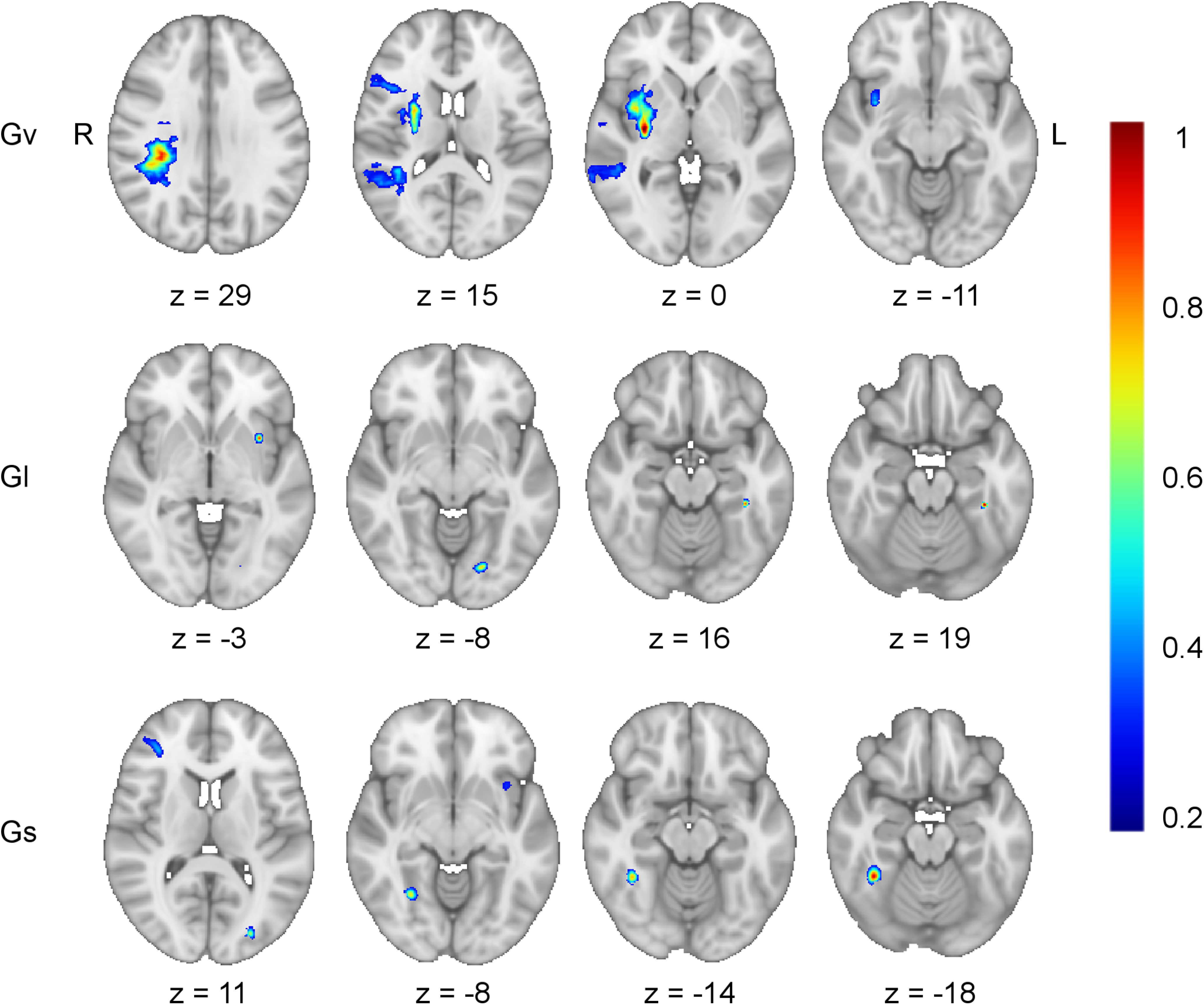
Bifactor model lesion-behavior mapping. Axial slices from the lesion-behavior maps estimated from the bifactor S-1 model illustrate the brain regions uniquely associated with each domain-specific cognitive ability after removing the variance attributable to general cognitive ability (g; see Fig. 2B). This included: visuospatial ability (Gv; r = 0.34, p < 0.001, optimal sparseness = −0.20), learning/memory (GI; r = 0.32, p < 0.001, optimal sparseness = −0.01), and processing speed (Gs; r = 0.19, p < 0.001, optimal sparseness = 0.08). The map for crystalized intelligence (Gc; r = 0.19, p < 0.001, optimal sparseness = 0.77) did not return any regions associated with impairment in this domain. Working memory (Gwm) demonstrated no remaining reliable variance after accounting for g and so was not depicted. For each map, the left hemisphere temporo-parietal white matter region implicated as important for g/Gwm (compare with Fig. 3) was no longer present in any of these maps. Voxel weights were derived from the optimized SCCAN and were scaled to values between 0 and 1.