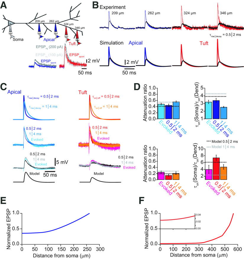
Figure 8.
The model predicts EPSP propagation in tuft dendrites. A, Schematic of recording configurations. Dual whole-cell recordings were performed in different cells at the indicated distances. Recordings from the apical dendrite are shown in blue, and recordings from tuft dendrites beyond the main bifurcation are shown in red. In the inset examples of spontaneous EPSPs from apical dendrite (blue) and tuft dendrite (red) are compared with EPSP-like current injections (τrise = 0.5 ms, τdecay = 2 ms) with amplitudes of 100 pA (gray) and 200 pA (black), suggesting that spontaneous EPSPs can be approximated by fast current injections. B, Comparison of measured somatic (black) and dendritic EPSPs evoked by EPSP-like current injections and the corresponding responses simulated in the model. Good agreement between experiment and simulation was found for both apical dendrite and distal tuft dendrite recordings in terms of local EPSP size and propagation toward the soma. C, Comparison of synaptically evoked EPSPs recorded in the soma and simultaneously in either the apical dendrite (left traces) or tuft (right traces) with EPSP-like current injections with different kinetics. Synaptically evoked EPSPs are well described by fast current injections. Modeling traces are shown after propagation to the soma. D, Bar graphs of average attenuation and change in EPSP slope for evoked EPSPs and EPSP-like current injections (τrise = 0.5 ms, τdecay = 2 ms; and τrise = 1 ms, τdecay = 4 ms, respectively). Dashed lines indicate the corresponding values obtained from the model. E, Plot of normalized EPSP amplitude as a function of distance from the soma for a modeled EPSP (τrise = 0.5 ms, τdecay = 2 ms) originating in the apical dendrite at the main bifurcation. F, Plot of normalized EPSP amplitude as a function of distance from the soma for a modeled EPSP (τrise = 0.5 ms, τdecay = 2 ms) originating in the distal part of the apical tuft. Most attenuation occurs within the tuft dendrite. Inset shows the evolution of the EPSP amplitude in the main apical dendrite on an expanded scale. Note that the EPSP, once it reaches the apical dendrite, is only little attenuated.

