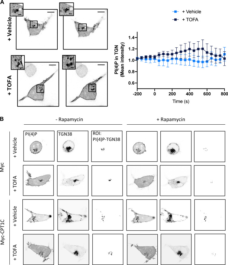
Figure S5.
Rapamycin-induced ER-TGN contacts. (A) No artificial contacts were formed without the addition of rapamycin. HEK cells were transfected and treated as in Fig. 8 A. PI(4)P levels in the TGN were live imaged with confocal microscopy. TOFA addition in these conditions does not have any effect. Data represent the mean ± SD from one experiment performed by biological replicates (n = 5 or 6 cells per condition). Scale bars = 7 µm; scale bars of inset magnifications = 1.75 µm. (B) Effect of CPT1C expression and TOFA treatment on PI(4)P levels at the TGN after induction of artificial ER-TGN contacts. HEK cells were transfected with mCherry-pHR-TcRb-FKBP and CFP-TGN38-FRB plasmids to induce artificial ER-TGN by rapamycin (5 µM, 30 min). PI(4)P was labeled with the lipid probe YFP-P4M. Cells were treated for 1 h with TOFA or vehicle 24 h after transfection. Cells were live imaged with confocal microscopy. PI(4)P, TGN, and the ROI of PI(4)P inside the TGN are shown. Individual cells are the same as the ones analyzed in Fig. 8 A.

