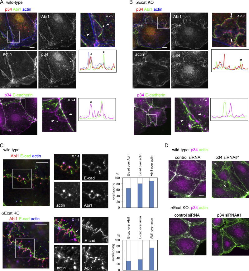
Figure 4.
Arp2/3 complex at junctions and its interaction with WRC and F-actin. (A and B) Coimmunostaining for p34/ARPC2 (p34), Abi1 and actin (top), and p34/ARPC2 and E-cadherin (bottom) in wild-type (A) or αEcat KO (B) Caco2 cells. J, the position of junction. Marginal or marginal plus submarginal cells are shown throughout this figure. (C) Coimmunostaining for Abi1, E-cadherin, and actin in wild-type or αE-cat KO Caco2 cells, which were incubated with 10 µM latrunculin A for 60 min. The boxed regions are enlarged. The overlapping ratios of these molecules were quantified. Magenta arrows, Abi1–actin coclusters; white arrows, Abi1–E-cadherin–actin coclusters; green arrows, E-cadherin fragments that do not overlap with other molecules. (D) Effect of p34/ARPC2 siRNA treatment on actin assembly. Arrows point to actin-positive structures that are not closely associated with the junctions. Magnification is shown in the partly enlarged images. Scale bars, 10 µm.

