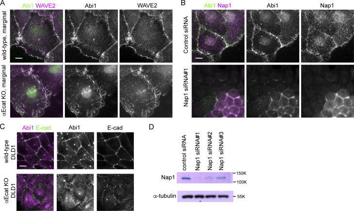
Figure S3.
Detection of WRC components in Caco2 and DLD1 cells. (A) Coimmunostaining for Abi1 and WAVE2 in wild-type or αEcat KO Caco2 cells. (B) Coimmunostaining for Abi1 and Nap1 in wild-type Caco2 cells that were treated with siRNA for Nap1. Junctional Abi1 was removed as a result of Nap1 depletion, whereas centrosomal staining with anti-Abi1 antibody was not, which suggests that this staining is due to nonspecific binding of the antibody to centrosomes. Consistently, antibodies for WAVE2 or Nap2 did not detect centrosomes. (C) Coimmunostaining for Abi1 and E-cadherin in wild-type and αEcat KO DLD1 cells. (D) Western blots for Nap1 in Nap1 siRNA-treated cells. Scale bars, 10 µm.

