Figure S2.
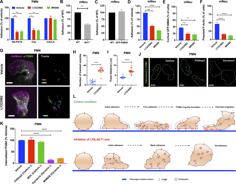
LTB4 signaling promotes the adhesion of primary human and mouse neutrophils in vitro. (A) PMNs were pretreated with the vehicle or 2 µM MK886 or 20 µM LY223982 for 20 min and stimulated with 25 nM fNLFNYK or 250 ng/ml C5a or 250 ng/ml CXCL8 for 15 min. Adhesion was calculated as described in the Materials and methods section and presented as a percentage relative to the respective vehicle controls for each chemoattractant. Data are plotted as mean ± SEM from n = 4 independent experiments. Two-way ANOVA using Dunnett’s multiple comparisons test was performed to determine statistical significance. (B and C) Neutrophils from WT (B and C), Blt1−/− (B), and GFP-NMIIA (C) mice were stimulated with 25 nM WKYMVm for 15 min on a fibrinogen-coated surface. Adhesion was calculated as described in the Materials and methods section and presented as a percentage relative to the WT controls. Data are plotted as mean ± SEM from n = 3 independent experiments in each panel. An unpaired t test with Welch’s correction was used to determine statistical significance, and no significant difference was observed between conditions in C. mNeu label stands for data from mouse primary neutrophils. (D–F) GFP-NMIIA neutrophils were pretreated with the vehicle or 2 µM MK886 or 20 µM LY223982 for 20 min, stimulated with 25 nM WKYMVm for 15 min, washed, fixed, and stained with rhodamine phalloidin. Adhesion was calculated as described in the Materials and methods section and presented as a percentage of vehicle-treated controls (D). Quantification of the percentage of neutrophils exhibiting cortical NMIIA (E) and polarized F-actin (F) in response to the above-mentioned treatments. Data are plotted as mean ± SEM from n = 3 independent experiments. One-way ANOVA using Dunnett’s multiple comparisons test was performed to determine statistical significance. mNeu label stands for data from mouse primary neutrophils. (G–I) PMNs were pretreated with either the vehicle or 20 µM LY223982 for 20 min. They were incubated with an Alexa Fluor 488–conjugated antibody against human ITGB2 (αITGB2, CTB104 clone; green) in combination with CellMask Deep Red (magenta) before stimulation with 100 nM fNLFNYK for 10 min on a fibrinogen-coated surface and acquired in 3D using time-lapse mode in confocal microscopy. The trajectory of the internalized vesicles was determined as described in the Materials and methods section (G). Tracks of individual ITGB2-containing vesicles between 5 and 7 min after stimulation are overlaid on the maximum-intensity projections derived from Z-stacks. Scale bars = 5 µm. See Video 7. Quantification of the number (H) and distance (I) traveled by ITGB2-containing vesicles for each condition. Data are plotted for individual cells with n = 16 cells in vehicle and n = 18 cells in LY223982 treatment from n = 3 independent experiments. An unpaired t test with Welch’s correction was used to determine statistical significance. (J and K) PMNs were pretreated for 20 min with vehicle (DMSO) or the indicated inhibitors of clathrin and dynamin (5 µM PitStop2 or 100 µM MDC or 50 µM dynasore or 2.5 µM MitMAB), stimulated for 10 min with 25 nM fNLFNYK, fixed, and imaged by confocal microscopy. Representative confocal images of PMNs with internalized ITGB2 under indicated conditions are presented (J). White dashed lines indicate cell boundary. Scale bars = 5 µm. The extent of ITGB2 internalization was determined as described in the Materials and methods section, and the data are plotted as the percentage change with respect to the vehicle (K). Data are represented as mean ± SEM from n = 3 independent experiments. One-way ANOVA using Dunnett’s multiple comparisons test was performed to determine statistical significance. (L) A model of ITGB2 trafficking regulated by the LTB4–BLT1 axis in PMNs. The figure depicts the progression of PMN adhesion on a fibrinogen-coated surface. In control condition, ITGB2 clusters and migrates to the bottom of the cell to form a ringlike structure, which then disassembles as the PMN begins to migrate. However, upon blockade of LTB4 sensing via BLT1, clusters of ITGB2 rapidly internalize and fail to assemble into a ringlike structure at the bottom of the PMN, resulting possibly in the de-adhesion of PMN with time. *, P < 0.05; **, P < 0.01; ***, P < 0.001; ****, P < 0.0001.

