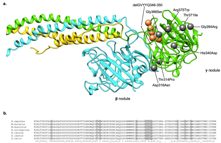
Figure 2.
Localization and conservation of HHHS -causing mutations in the fibrinogen γ nodule. (a) The localization of all the mutations responsible for the HHHS phenotype, described in the literature, are indicated as grey spheres (in case of missense mutations), and orange spheres (in case of deletion). Mutations are numbered on the mature protein. The fibrinogen chains (Aα, Bβ, γ) are represented in yellow, cyan, and green, respectively. The model was obtained using the pdb structure 3GHG, and the UCSF Chimera package. (b) Multiple alignments of the fibrinogen γ chain terminal regions of different species. Sequences were retrieved from NCBI, and aligned using clustalW. The last line of the alignment shows in a schematic way the conservation among species. The * symbol indicates a conserved residue, the : symbol indicates conservation between groups of strongly similar properties, the . symbol indicates conservation between groups of weakly similar properties, the space indicates lack of conservation. Amino acids shaded in grey corresponds to those involved in HHHS mutations.

