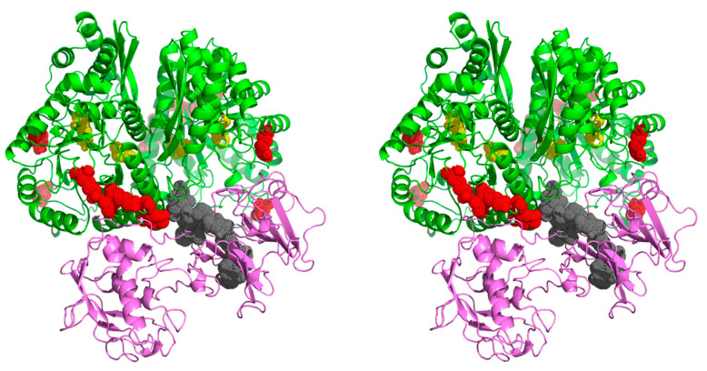
Figure 5.
Proposed model of the interaction between C. albicans enolase (green) and human VTR (purple). The docking was performed for the C. albicans enolase dimeric structure (template PDB ID: 1EBH [58]) and VTR structure obtained based on multiple templates (PDB ID: 3C7X, 1OC0, 2JQ8 and 3BT1 [61,62,63,64]). The enolase peptides and the VTR peptide identified in the chemical mapping experiments are indicated in red and gray, respectively. The active site residues of the catalytic center of enolase are indicated in yellow. The 3D molecular model is presented in a wall-eyed stereo view. An additional file Enolase_VTR.mpg with the PyMOL-generated video presentation of this model is attached to the article together with the Supplementary Materials (Video S1).

