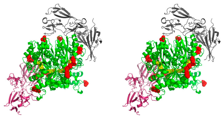
Figure 6.
Stereo image (wall-eyed) of the interaction between C. albicans enolase and human FN. The docking was performed for the C. albicans enolase dimeric structure (template PDB ID: 1EBH [58]) and FN fragment (PDB ID: 3M7P [60]). The two models for the protein-protein interaction between a human FN fragment (black and pink) and C. albicans enolase (green with active site residues of the catalytic center indicated in yellow) were generated using ClusPro 2.0: protein-protein docking software. The enolase peptides identified in the chemical mapping experiments are marked in red. Additional files Enolase_FN_1.mpg and Enolase_FN_2.mpg with the PyMOL-generated video presentations of these models are attached to the article together with the Supplementary Materials (Videos S2 and S3).

