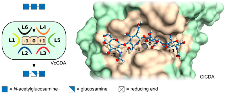
Figure 2.
Fungal chitin deacetylases (CDA) (right), which are better known than e.g., bacterial (left) or insect CDAs, typically possess four subsite binding sites (ranging from {–2} to {+1}) within a substrate binding cleft, highlighted in beige, each of which binds one monosaccharide subunit of the substrate, a chitin or chitosan oligomer or polymer. Each subsite can have its own specificity or preference for binding a N-acetylglucosamine or a glucosamine unit. The substrate binding cleft, shown in beige, may be delineated by six protein loops (L1 to L6) which may be flexible, allowing an induced fit of the enzyme. Left, schematic representation of the bacterial VcCDA from Vibrio for which the surrounding loops where first described; right, 3D-model of the fungal ClCDA from the Colletotrichum with bound substrate, chitin tetraose.

