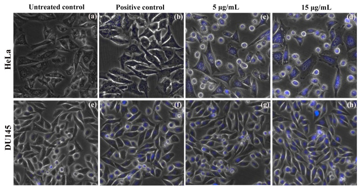
Figure 7.
Nuclear staining using DAPI on HeLa and DU145 carcinoma cells observed at 20× magnification under fluorescent inverted microcope. Untreated negative controls: (a) HeLa, (e) DU145. 20 µg/mL leaf extract taken as positive controls for (b) HeLa and 15 µg/mL for (f) DU145. ZnO NPs treatment 5 µg/mL: (c) HeLa, (g) DU145; and 15 µg/mL ZnO NPs treatment: (d) HeLa, (h) DU145 cell lines.

