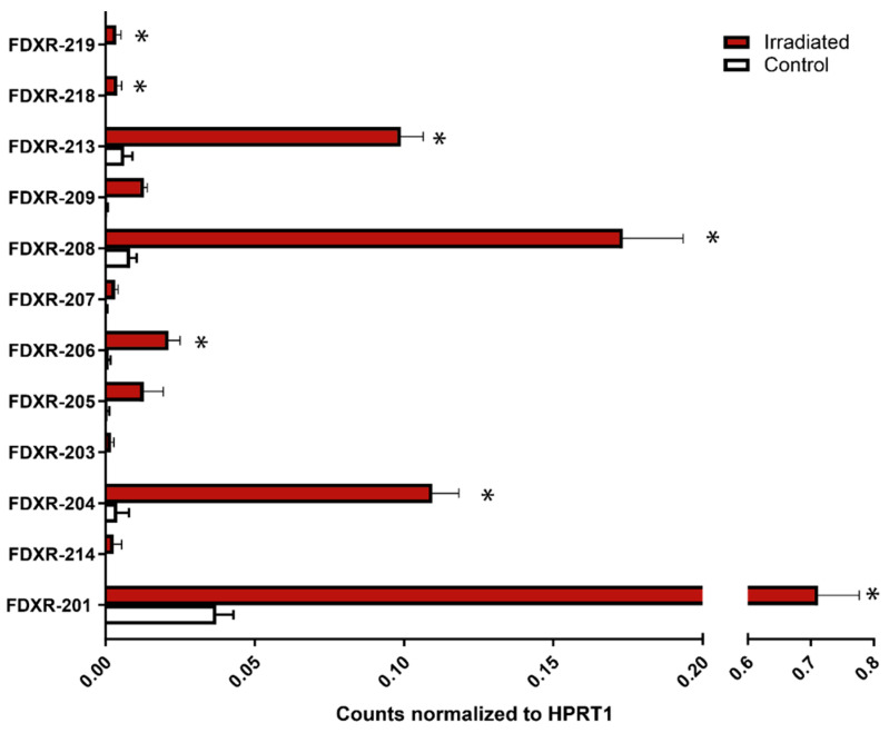
Figure 6.
Variants identified by nanopore sequencing. Blood samples were expose to a 2 Gy dose (0.5 Gy/min) and incubated at 37 °C for 24 h. RNA was extracted and poly A+ enriched before preparing the library using a cDNA-PCR kit (PCS109). The sequencing was performed in a PromethION (Oxford Nanopore Technologies). Counts were normalized by HPRT1. Data are shown as mean ± SEM (n = 3). Statistical analyses were performed in log-transformed data. Significant differences (paired t-test, p ≤ 0.05) with the control were indicated with an asterisk (*).

