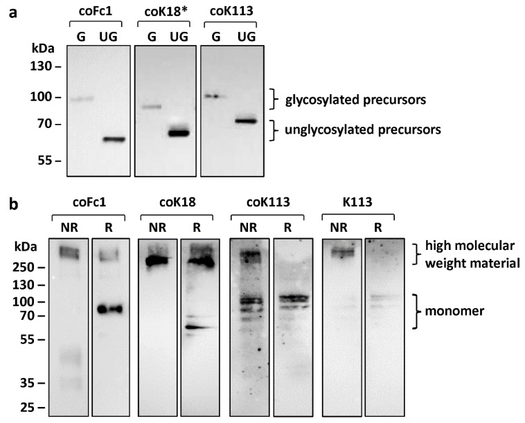
Figure 2.
Western blot analysis for characterization of post-translational modifications of HERV ENV. (a) Detection of glycosylated (G) or unglycosylated (UG) envelope proteins in cell lysates of transiently transfected HEK293 cells. Notice the shift in protein size after deglycosylation by PNGase F. (b) Higher molecular weight material resulting from expression of HERV ENV precursors via intramolecular disulfide bridges analyzed by comparison of non-reducing (NR) and reducing (R) sample buffer conditions. The monomers are only visible when reducing (R) sample buffer was used. Either anti-FLAG antibody (coFc1) or anti-HERV-K SU HERM1821-5 antibody (coK18*, coK113) was used for detection of ENV.

