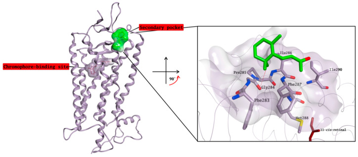
Figure 11.
Crystal structure of β-ionone (carbon atoms in green) in complex with rhodopsin in which the chromophore-binding pocket is already occupied by 11-cis-retinal (carbon atoms in garnet). β-ionone is bound to a small, surface-exposed and highly hydrophobic pocket. The binding site area is represented as the molecular surface. Rhodopsin is represented as the lilac ribbon. On the binding site cut out rhodopsin is represented as a lilac tube for clarity.

