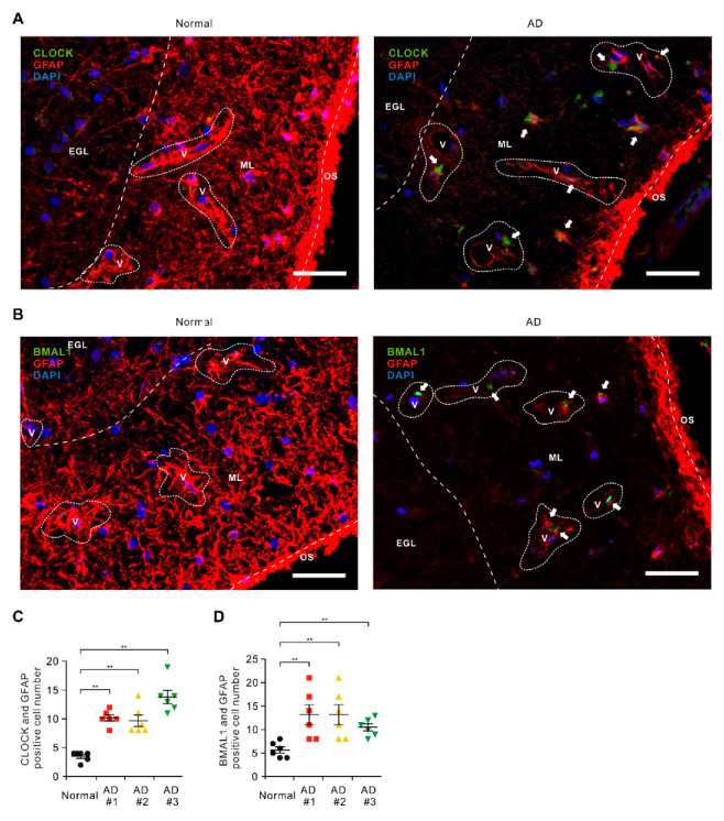
Figure 1.
The levels of circadian locomotor output cycles kaput (CLOCK) and brain muscle ARNT-like1 (BMAL1) are elevated in impaired astrocytes in cerebral cortex region from patients with Alzheimer’s disease (AD). (A) Representative immunofluorescence images of CLOCK expression in cerebral cortex region from patients with AD (AD) or non-AD (normal) showing CLOCK (green) in astrocytes expressing the astrocytes marker GFAP (red) around blood vessels (V) (n = 3 per group, n = 6 images per individual subject). DAPI-stained nuclei are shown in blue. Outer surface (OS); molecular layer (ML); external granular layer (EGL). Scale bars, 20 μm. (B) Representative immunofluorescence images of BMAL1 expression in cerebral cortex region from patients with AD (AD) or non-AD (normal) showing BMAL1 (green) in astrocytes expressing the astrocytes’ marker GFAP (red) around blood vessels (V) (n = 3 per group, n = 6 images per individual subject). DAPI-stained nuclei are shown in blue. Outer surface (OS); molecular layer (ML); external granular layer (EGL). Scale bars, 20 μm. DAPI-stained nuclei are shown in blue. (C) Quantification of CLOCK positive astrocytes from immunofluorescence images in cerebral cortex region from patients with AD (AD) or non-AD (normal) (n = 3 per group, n = 6 images per individual subject). Data are mean ± standard deviation (SD). **, p < 0.01 by Student’s two-tailed t-test. (D) Quantification of BMAL1 positive astrocytes from immunofluorescence images in cerebral cortex region from patients with AD (AD) or non-AD (normal) (n = 3 per group, n = 6 images per individual subject). Data are mean ± standard deviation (SD). Symbols, which are expressed by white dotted line, indicate the outline of BBB. **, p < 0.01 by Student’s two-tailed t-test.

