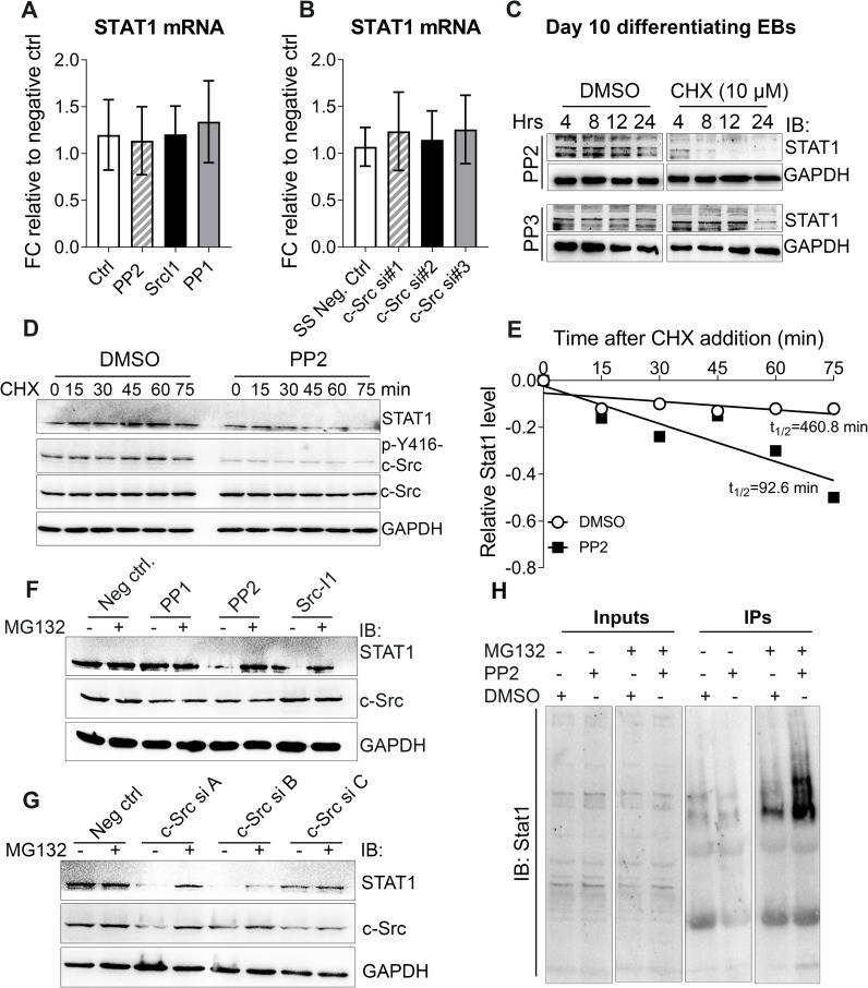
Fig 4. c-Src enhances STAT1 protein stability.
(A and B) Real time qPCR analysis of STAT1 expression upon pharmacological inhibition of c-Src using 10 μM of PP1, SrcI-1, and PP2 for 2 hours where DMSO served as negative control (A) and c-Src downregulation using with three different distinct siRNAs where silencer™ select negative siRNA served as control for 48 hours (B) in MC3T3E1 cells. Data shown represent the means (± SD) of triplicates. (C) c-Src inhibition and its effect on STAT1 protein level in mESCs. Differentiating EBs were exposed to 10 μM of PP2 or PP3 for 24 hours prior to treatment with CHX (10 μM) or DMSO. Lysates were collected at the indicated times and subjected to WB analysis using STAT1 antibody. GAPDH served as loading control. (D) Inhibition of c-Src activity and its effect on STAT1 half-life in MC3T3-E1 cells. Cells were subjected to PP2 (10 μM) or DMSO (solvent ctrl) treatments for 2 hrs, prior to addition of CHX (10 uM). Harvested cells at the indicated time points were subjected to WB analysis to examine STAT1 expression. GAPDH served as a loading control. (E) Quantification of STAT1 stability assays. STAT1 band densities were quantified, first normalized to the corresponding GAPDH and then to t = 0 DMSO controls. (F and G) Inhibition of proteasomal degradation by MG132 and its effect on STAT1 loss resulted from c-Src inhibition. MC3T3-E1s were treated with three different c-Src inhibitors including PP2, Src I-1, and PP1 (10 μM) for 24 hrs (F) or were transfected with control siRNA or different combinations of three different specific c-Src siRNA for 48 hrs (A = c-Src siRNAs #1+ #2, B = c-Src siRNAs #1+ #3, and C = c-Src siRNAs #2+ #3) (G). Before harvest, cells were treated with MG132 (10 μM) for 4 hrs as indicated. STAT1, c-Src, and p-c-Src Y416 protein levels were analyzed by immunoblots, with GAPDH as a loading control. (H) Inhibition of c-Src activity and its effect on STAT1 ubiquitination. Proteasomal degradation was inhibited by MG132 in MC3T3-E1 cells for two hours. DMSO served as the negative control. After two hours treated cells underwent second treatments with PP2 (10 μM) or DMSO for another 2 hours. Lysates were subjected to IP assay using STAT1 antibody. Precipitated immunocomplexes were then analyzed by WB using both STAT1 antibody.

