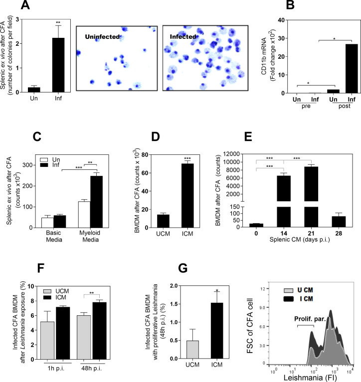
Fig 2. The infected environment in VL induces the proliferation of myeloid cells.
A, Number of colonies in a colony formation assay (CFA) using splenic myeloid cells from uninfected hamsters (Un) or from hamsters infected with L. donovani (Inf). Colonies were counted on the inverted microscope after 12 days of incubation (**p<0.008, Mann-Whitney Test); Right, representative Giemsa staining of cells at day 12 of CFA, showing some bi-nucleated cells; B, Expression of the pan-myeloid CD11b marker before culture (pre) and at 12 days of CFA (post), determined by qPCR (*p<0.05, Kruskal-Wallis Test); C, Number of cells at 12 days of CFA with splenic myeloid cells exposed to a basic medium (without myeloid growth factors) or complete medium (with myeloid growth factors). The number of cells was determined by Cell Titer Glo (**p<0.01; ***p<0.0001, Kruskal-Wallis Test); D, Number of bone marrow derived cells (BMDM) at 12 days of exposure to a CFA with CM from spleen of uninfected (UCM) or infected hamsters (ICM) (***p<0.0001, Unpaired T test); E, Number of BMDM at 12 days of CFA exposed to splenic CM obtained from spleens of uninfected hamsters (time 0) or from hamsters infected with L. donovani at the indicated times post-infection (14–28 days p.i.) ***p<0.001, Tukey-Kramer Multiple Comparisons Test); F, Percentage of infected CFA cells after in vitro exposure to L. donovani promastigotes (1h or 48h of in vitro infection). CFA cells obtained after 12 days of BMDM exposed to UCM or ICM as determined by flow cytometry (**p = 0.03, Unpaired t test); G, Percentage of cells from CFA with proliferative parasites after 48h of in vitro infection with L. donovani promastigotes. Parasites were prelabeled with a fluorescent proliferation tracer before in vitro infection (*p< 0.004, Unpaired T test). Right, representative histogram showing fluorescence intensity of proliferative parasites (with diluted fluorescence) compared with parental parasites.

