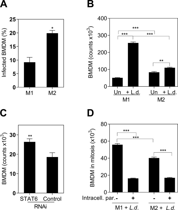
Fig 3. The proliferative capacity of BMDM was limited by intracellular L.donovani parasites and M2-polarization.
A, Proportion of infected M1 or M2 BMDM after 48 hours of exposure to L. donovani promastigotes (*p = 0.03, Unpaired t test, representative of 3 experiments); B, Absolute number of M1 or M2 BMDM stimulated with L. donovani promastigotes (+L.d.) or without parasite stimulation (Un). Number of cells counted by luminometry (*** p<0.001, **p<0.01, Tukey-Kramer Multiple Comparisons Test, representative of 3 different experiments); C, Number of BMDM after STAT6 silencing (STAT6 RNAi) compared with control silenced cells (control RNAi), 48h after exposure to L. donovani parasites (** p = 0.0079, Mann-Whitney Test). Data is the number of stimulated cells minus non-stimulated cells; D, Number of M1 or M2-polarized BMDM Ki-67+ (mitosis) containing intracellular parasites (+) or remaining free of intracellular parasites (-), after 48h of exposure to fluorescent-L. donovani (Number of cells in mitosis = percentage ki-67+ cells by flow cytometry x number of cells by luminometry/ 100) (*** p<0.0001, Tukey-Kramer Multiple Comparisons Test, representative of 3 different experiments).

