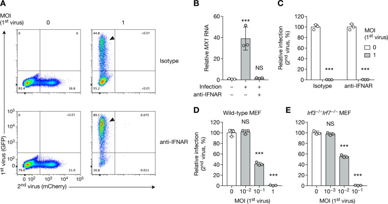
Fig 2. CHIKV SIE is independent on type I interferon.
(A–C) HFFs were treated with human IFNAR blocking antibody or isotype control at 5 μg/mL for 1 h, then were infected with CHIKV-GFP for 24 h at the indicated MOI, followed by CHIKV-mCherry at MOI 1 for 24 h. Blocking antibody treatment was maintained throughout the experiment. Representative flow cytometry plots (A) and quantification of infected cells (C) and are shown. Arrows highlight the difference in GFP infection between isotype control and blocking antibody treated samples. MX1 RNA levels were assessed by RT–qPCR (B). (D, E) WT (D) or Irf3−/−Irf7−/−MEF cells (E) were infected with CHIKV-GFP at the indicated MOI for 16 h, then with CHIKV-mCherry at MOI 5 (WT) or 3 (Irf3−/−Irf7−/−) for 8 h, and subsequently analyzed by flow cytometry. Bars indicate mean and SD of biological triplicates, and data are representative of at least two independent experiments. NS, not significant; ***p < 0.001 (one-way analysis of variance followed by Dunnett’s post-test).

