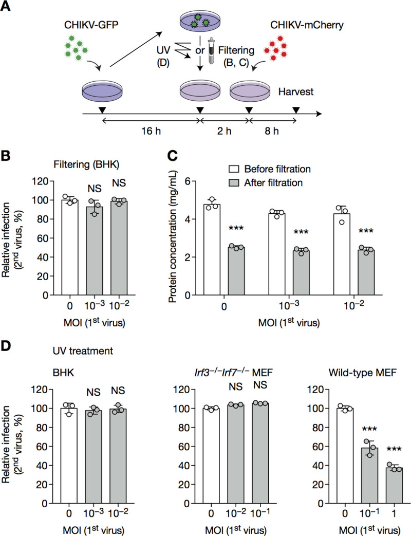
Fig 3. CHIKV SIE is a cell-intrinsic mechanism.

BHK cells, WT or Irf3−/−Irf7−/−MEF cells were infected with CHIKV-GFP at the indicated MOI (A), then supernatant was subject to filtering (B, C) or UV irradiation (D) and overlaid onto fresh cells, which were challenged 2 h later with CHIKV-mCherry at an MOI of 1 (BHK), 5 (wild-type MEF) or 3 (Irf3−/−Irf7−/− MEF) for 8 h, then harvested and analyzed by flow cytometry. Protein concentration before and after filtration was assessed in C. Bars indicate mean ± SD of biological triplicates, and data are representative of two independent experiments. NS, not significant; ***p < 0.001 (one-way analysis of variance followed by Dunnett’s post-test).
