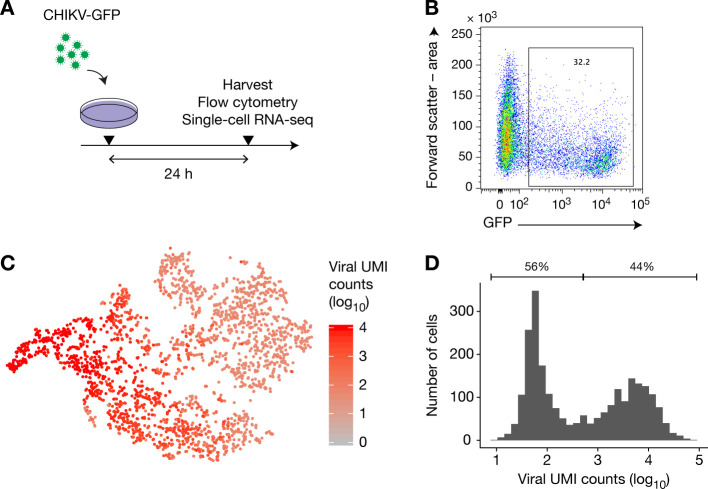
Fig 4. After primary infection, all cells contain viral RNA.
(A) HFFs were infected with CHIKV-GFP at MOI 1 for 24 h, then harvested and separated into two samples for analysis by flow cytometry and single-cell RNA-sequencing using 10x genomics Single Cell 3’. (B) Flow cytometry analysis of the infected cells. (C) T-SNE analysis of the RNA-sequencing data. (D) Distribution of the number of CHIKV unique molecular identifiers (UMI) per cell.

