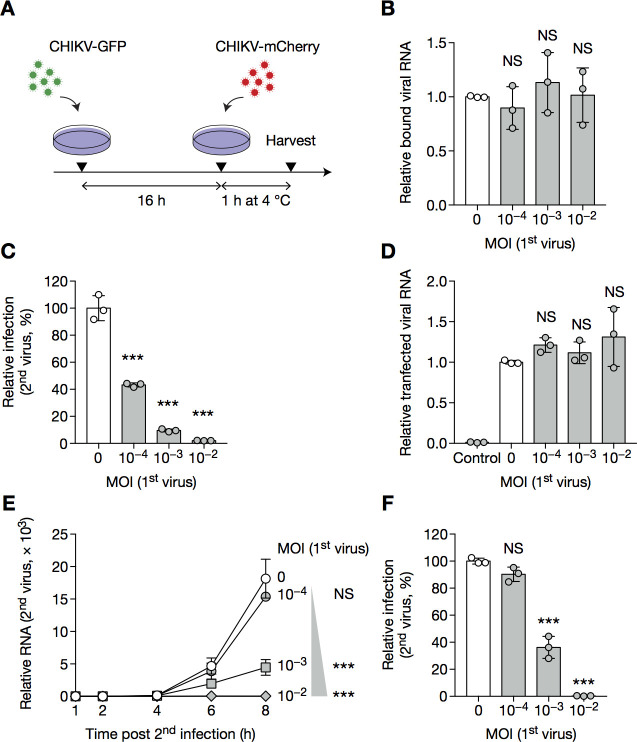
Fig 7. Primary virus inhibits replication of the challenge virus.
(A–B) BHK cells were infected with CHIKV-GFP for 16 h at the indicated MOI, then with CHIKV-mCherry at MOI 1 for 1 h at 4°C and harvested for RT–qPCR specific for mCherry. (C, D) BHK cells were infected with CHIKV-GFP for 16 h at the indicated MOI, then transfected with in vitro transcribed RNA coding for CHIKV-mCherry. 12 h post-transfection, cells were harvested and analyzed by flow cytometry (C); 4 h post-transfection, transfection efficiency was controlled by RT–qPCR (D). Control wells were overlaid with transfection mix for 5 min at room temperature, to assess background noise after washing. (E, F) BHK cells were infected with CHIKV-GFP at the indication MOI for 16 h then CHIKV-mCherry at MOI 1 for the indicated time, and harvested for RT–qPCR specific for mCherry (E) or analyzed by flow cytometry (F) to provide the protein data for this particular day of experiment. Bars indicate mean ± SD of biological triplicates, and data are representative of at least two independent experiments. NS, not significant; *p < 0.05, **p < 0.01, ***p < 0.001 (one-way analysis of variance followed by Dunnett’s post-test).

