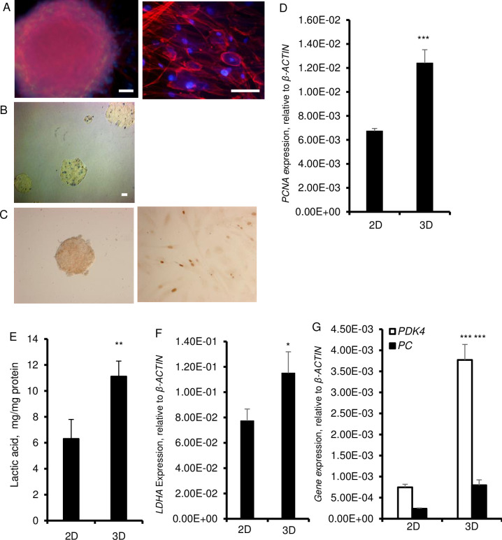
Fig 1. Cell morphology and metabolic features of fibroblasts grown in 3D.
HDF were cultured in 2D and in 3D cultures on ultra-low attachment plates for 6 days in αMEM supplemented with 20% Knockout Serum Replacement, 5 ng/ml basic FGF, 10 ng/ml EGF and with 0.1 mM nonessential amino acids (NEAA). A. Staining of cytoskeletal F-actin with phalloidin (red fluorescence) in spheroids and monolayer cultures. Nuclei are stained with Hoechst (blue fluorescence). Spheroid was photographed at 20× magnification (left) and monolayer cultured cells were photographed at 40× magnification (right). Scale bars = 200 μm. B. Staining of spheroids with trypan blue. C. BrdU staining (brown) in HDF spheroids (left) and in 2D culture after 6 days. D. Expression of proliferating cell nuclear antigen (PCNA) at the mRNA level. β-ACTIN was used as a housekeeping control. E. Amount of lactic acid in supernatant over a 6-day culture period. F. Expression of LDHA mRNA. β-ACTIN was used as a as a housekeeping control. G. Expression of PDK4 and PC mRNA. β-ACTIN was used as a housekeeping control. Data are shown as means ± SD from 3 independent experiments. Statistical significance is shown as * (p≤0.05), ** (p ≤0.005), and *** (p ≤0.0005) in 3D as compared to 2D cultured cells.

