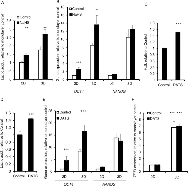
Fig 4. Effect of H2S donors, NaHS and DATS on lactic acid production and expression of OCT4 and NANOG in 3D spheroids.
A. Extracellular lactic acid in presence of 50 μM NaHS after 6 days. B. qPCR of OCT4 and NANOG in presence of 100 μM NaHS for 6 days. C-G. HDFs were cultured for 6 days in in 2D and 3D in the absence and the presence of 50 μM DATS. C. Extracellular H2S in presence of 50 μM DATS after 6 days. D. Extracellular lactic acid in presence of 50 μM DATS after 6 days. E. qPCR of OCT4 and NANOG. β-ACTIN was used as a housekeeping control. F. qPCR of TET1. Data are presented as the mean ± SD from 3–6 independent experiments. β-ACTIN was used as a housekeeping control. Statistical significance is shown as * (p≤0.05), ** (p ≤0.005), and *** (p ≤0.0005) in 3D as compared to 2D cultured cells.

