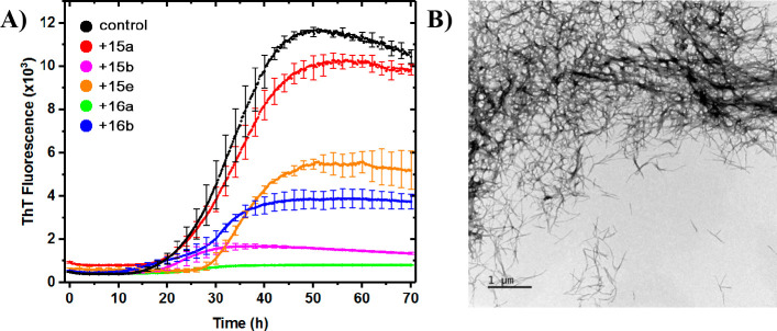
Figure 2.
(A) Representative selection of ThT fluorescence traces. Each experiment was performed in triplicate (Figure S1) and normalized to the average. Compounds 15b and 16a appear to bind fibrils strongly, whereas 15a does not seem to bind to any significant extent. 15e and 16b are borderline. (B) TEM picture of fibrils formed in the presence of compound 16a (green trace).

