
Figure 9. MFAP4 deficiency alleviates aortic banding‐induced ventricular arrhythmia.
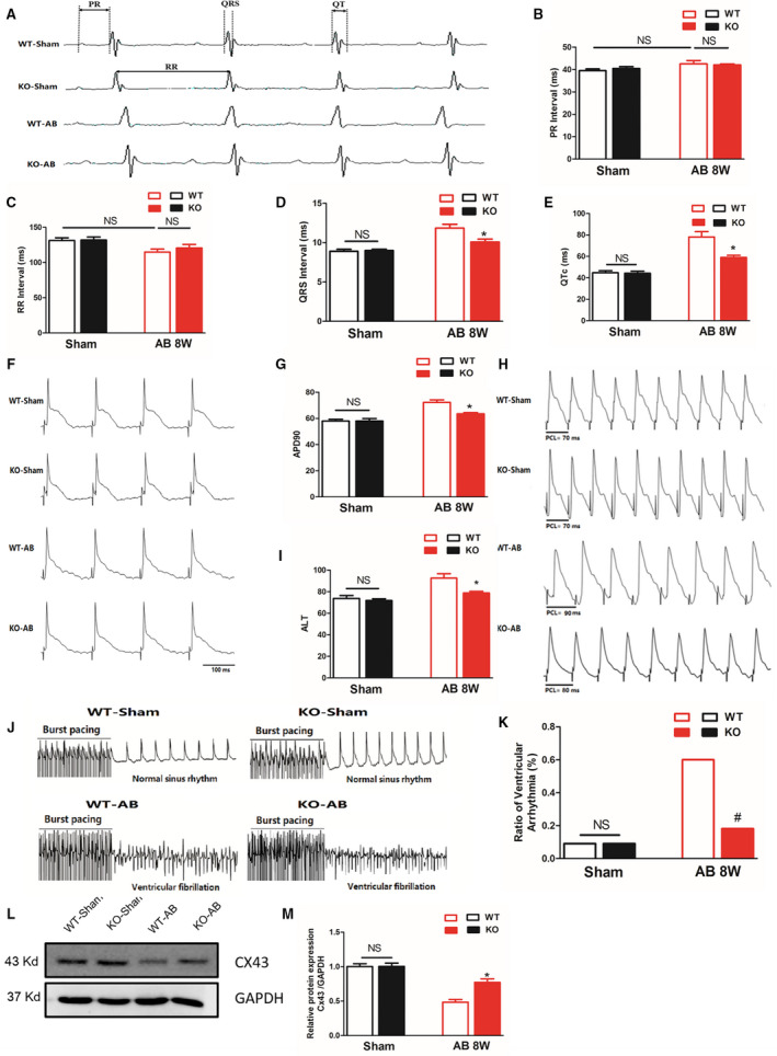
A, Representative ECG recordings and a comparison of ECG parameters. B through E, Statistical analysis of the PR, RR, QRS, and QTc intervals in MFAP4 (microfibrillar‐associated protein 4)‐knockout (KO) and wild‐type (WT) mice 8 weeks after the sham operation or aortic banding (AB) surgery (n=8 for the WT‐sham and KO‐sham groups; n=13 for the WT‐AB group; n=7 for the knockout [KO]‐AB group). F and G, Representative action potential figures and statistical analysis of the APD90 values in MFAP4‐KO and WT mice 8 weeks after the sham operation or AB surgery (n=8). H and I, Representative electric alternans figures and statistical analysis of the ALT thresholds in MFAP4‐KO and WT mice 8 weeks after the sham operation or AB surgery (n=8). J and K, Representative arrhythmia induced by burst‐pacing stimulation and statistical analysis of MFAP4‐KO and WT mice 8 weeks after the sham operation or AB surgery (n=11 for the WT‐sham, KO‐sham, and KO‐AB groups; n=10 for the WT‐AB group). L and M, Representative western blots, and statistical analysis of electroconduction‐related protein (Connexin 43) levels in MFAP4‐KO and WT mouse heart tissues (n=4). AB indicate aortic banding; AB 8W, 8‐week aortic banding group; ALT, action potential duration alternans; APD90, 90% action potential duration; Cx43, Connexin 43; HW/BW, heart weight/body weight; KO, knockout mice; MFAP4, microfibrillar‐associated protein 4; and WT, wild‐type. *P<0.05 represents the comparison between WT‐AB and KO‐AB group.